Leg Bone Diagram : Infographic Diagram Of Human Femur Bone Or Leg Bone ...
Leg Bone Diagram : Infographic Diagram Of Human Femur Bone Or Leg Bone .... The major bones of the leg are the femur (thigh bone), tibia (shin bone), and adjacent fibula, and these are all long bones.the patella (kneecap) is the sesamoid bone in front of the knee.most of the leg skeleton has bony prominences and margins that can be palpated and some serve as anatomical landmarks that define the extent of the leg. Depending on the origin of the discomfort, upper leg pain symptoms can be a chronic nuisance or acute and debilitating. Any disorder or defect in the knee bone can restrict the activities of the leg which can directly affect our locomotion. The photos you provided may be used to improve bing image processing services. To explain the term in layman's language, it is the heel bone in the skeletal system.
ads/bitcoin1.txt
The tibia and fibula are two long bones that run parallel to each other, forming the scaffold of the leg and providing attachment points for many muscles. The smaller lateral bone of the lower leg. The major bones of the leg are the femur (thigh bone), tibia (shin bone), and adjacent fibula, and these are all long bones.the patella (kneecap) is the sesamoid bone in front of the knee.most of the leg skeleton has bony prominences and margins that can be palpated and some serve as anatomical landmarks that define the extent of the leg. This area is commonly referred to as the calf. The tibia, commonly known as the 'shin bone', is the largest and most medial of the two.you can palpate its anterior border when you run your finger down the anterior aspect of your leg.
Osteology Unit - M.Y. Online Portfolio from mkyousif17.weebly.com Learn vocabulary, terms, and more with flashcards, games, and other study tools. Long bones, short bones, flat bones, and irregular bones.) long bones are longer than they are wide, with spongy bones at both ends and a cavity filled with bone marrow in the shaft. The lower leg extends from the knee to the ankle. The muscles in the upper leg power many of our movements. With different grades of sprains depending on severity. The bones together make up the hip. There are in all 7 bones, which fall under tarsal bones category. This area is commonly referred to as the calf.
Long bones have a thick outside layer of compact bone and an inner medullary cavity containing bone marrow.
ads/bitcoin2.txt
The foot bones shown in this diagram are the talus, navicular, cuneiform, cuboid, metatarsals and calcaneus. The rounded, proximal end is the head of the femur, which articulates with the acetabulum of the hip bone to form the hip joint. The lower leg is comprised of two bones, the tibia and the smaller fibula. Learn vocabulary, terms, and more with flashcards, games, and other study tools. Learn vocabulary, terms, and more with flashcards, games, and other study tools. It is the largest bone in the body and is the only bone in the upper leg. Below given knee diagram will help you to understand the various parts and functioning of the knee joint. Human anatomy for muscle, reproductive, and skeleton. A long bone has a shaft and 2 ends. Start studying lab test 2: The tibia, commonly known as the 'shin bone', is the largest and most medial of the two.you can palpate its anterior border when you run your finger down the anterior aspect of your leg. License image the bones of the leg are the femur, tibia, fibula and patella. The bones of the leg and foot form part of the appendicular skeleton that supports the many muscles of the lower limbs.
Below given knee diagram will help you to understand the various parts and functioning of the knee joint. Bone structure of leg, above and below The tibia and fibula are two long bones that run parallel to each other, forming the scaffold of the leg and providing attachment points for many muscles. Depending on the origin of the discomfort, upper leg pain symptoms can be a chronic nuisance or acute and debilitating. The major bones of the leg are the femur (thigh bone), tibia (shin bone), and adjacent fibula, and these are all long bones.the patella (kneecap) is the sesamoid bone in front of the knee.most of the leg skeleton has bony prominences and margins that can be palpated and some serve as anatomical landmarks that define the extent of the leg.
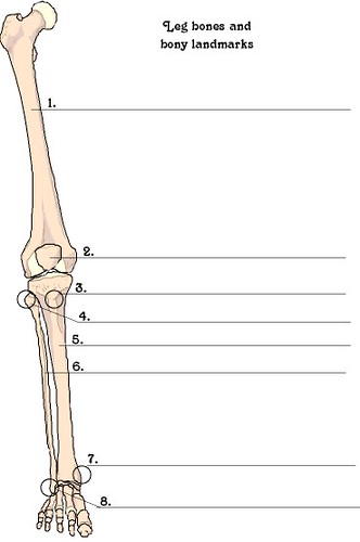
leg bones and bony landmarks | Lorie Warren | Flickr from c2.staticflickr.com The tibia and the fibula, at the top of the ankle joint. Structure of anatomy leg and foot 6 photos of the structure of anatomy leg and foot leg foot anatomy, leg foot bones, leg foot cramps, leg foot cramps at night, leg foot massage, leg foot numbness, leg foot pain, leg foot tattoos, foot, leg foot anatomy, leg foot. The lower leg is comprised of two bones, the tibia and the smaller fibula. To explain the term in layman's language, it is the heel bone in the skeletal system. At the same time, the bones and joints of the leg and foot must be strong enough to support the body's weight while remaining. The lower leg extends from the knee to the ankle. These landmarks are the anterior superior iliac spine. With different grades of sprains depending on severity.
The major bones of the leg are the femur (thigh bone), tibia (shin bone), and adjacent fibula, and these are all long bones.the patella (kneecap) is the sesamoid bone in front of the knee.most of the leg skeleton has bony prominences and margins that can be palpated and some serve as anatomical landmarks that define the extent of the leg.
ads/bitcoin2.txt
Muscles and tendons of the leg, find out more about muscles and tendons of the leg. The bones together make up the hip. The tibia, commonly known as the 'shin bone', is the largest and most medial of the two.you can palpate its anterior border when you run your finger down the anterior aspect of your leg. With different grades of sprains depending on severity. The knee joint is the largest joint in the body and is primarily a hinge joint, although some sliding and rotation occur. The diagram of bones in the ankle and foot is given below: The tibia and the fibula, at the top of the ankle joint. Any disorder or defect in the knee bone can restrict the activities of the leg which can directly affect our locomotion. Related posts of leg bones anatomy diagram structure of anatomy leg and foot. Leg bone anatomy diagram diagram of human leg human anatomy diagram 10 / 10 ( 1 vote ) in this image, you will find femur, medial epicondyle of the femur, patella, tibial tuberosity, anterior rest of the tibia, a medial surface of the tibia, lateral epicondyle of the femur, head of the fibula, fibula, medial malleolus of the tibia, lateral. The tarsal bones in the foot are located amongst tibia, metatarsal bones, and fibula. See more ideas about muscle anatomy, human anatomy and physiology, body anatomy. The lower leg is comprised of two bones, the tibia and the smaller fibula.
Back muscle diagram 12 photos of the back muscle diagram back and shoulder muscle diagram, back muscle diagram exercise, back muscle diagram pain, front and back. Learn vocabulary, terms, and more with flashcards, games, and other study tools. The knee joint is the largest joint in the body and is primarily a hinge joint, although some sliding and rotation occur. The tibia and fibula are two long bones that run parallel to each other, forming the scaffold of the leg and providing attachment points for many muscles. Start studying lab test 2:
16 best Bones in the Leg images on Pinterest | Human body ... from i.pinimg.com It is the largest bone in the body and is the only bone in the upper leg. (there are four types of bone: Learn vocabulary, terms, and more with flashcards, games, and other study tools. Start studying lab test 2: The lower leg extends from the knee to the ankle. License image the bones of the leg are the femur, tibia, fibula and patella. The femur, or thighbone, is the longest and largest bone in the human body. Related posts of diagram of leg bones human skeleton with each bone name.
The tibia, commonly known as the 'shin bone', is the largest and most medial of the two.you can palpate its anterior border when you run your finger down the anterior aspect of your leg.
ads/bitcoin2.txt
The upper leg, in particular, is comprised of bones and muscles that are susceptible to injury, particularly when excess strain is placed upon them. Start studying lab test 2: The foot bones shown in this diagram are the talus, navicular, cuneiform, cuboid, metatarsals and calcaneus. A long bone is a bone that has greater length than width. Below given knee diagram will help you to understand the various parts and functioning of the knee joint. Related posts of leg bones anatomy diagram structure of anatomy leg and foot. The femur is known as a long bone. The lower leg extends from the knee to the ankle. See more ideas about muscle anatomy, human anatomy and physiology, body anatomy. They support the legs to bear the body weight and also help in proper locomotion. These muscles work together to produce movements such as standing, walking, running, and jumping. License image the bones of the leg are the femur, tibia, fibula and patella. Also called the shin bone, the tibia is the longer of the two bones in the.
ads/bitcoin3.txt
ads/bitcoin4.txt
ads/bitcoin5.txt
0 Response to "Leg Bone Diagram : Infographic Diagram Of Human Femur Bone Or Leg Bone ..."


0 Response to "Leg Bone Diagram : Infographic Diagram Of Human Femur Bone Or Leg Bone ..."
Post a Comment